Wednesday 5 June 2013

First Dual-Action Compound Kills Cancer Cells, Stops Them from Spreading


June 5, 2013 — Scientists are reporting development and successful lab tests on the first potential drug to pack a lethal one-two punch against melanoma skin cancer cells. Hit number one destroys cells in the main tumor, and the second hit blocks the spread of the cancer to other sites in the body, according to their report in the journal ACS Chemical Biology.


Nathan Luedtke and colleagues explain that the spread of melanoma and other forms of cancer beyond the original location -- a process called metastasis -- makes cancer such a serious disease. Photodynamic therapy (PDT), which involves administering a drug that kills cancer cells when exposed to light, already is available. But PDT works only on the main tumor and has other drawbacks. Luedtke's team set out to find an improved approach to PDT.
The scientists describe successful tests in laboratory mice of one compound they synthesized that not only killed melanoma cells, but also stopped them from metastasizing by blocking a key signal inside the tumor cells. The compound "provides the first example of a preclinical candidate possessing both of these properties," the scientists state.
The authors acknowledge funding from the Swiss National Science Foundation.

Genetic Testing of Rare Blood Cancer Reveals New Mutation


June 5, 2013 — A recent article in the New England Journal of Medicine describes genetic testing of a rare blood cancer called atypical chronic neutrophilic leukemia (CNL) that revealed a new mutation present in most patients with the disease. The mutation also serves as an Achilles heel, allowing doctors at the University of Colorado Cancer Center to prescribe a never-before-used, targeted treatment. The first patient treated describes his best snowboarding season ever


 "I'm a crazy sports fan," says the patient. "I go 30 days a season. I may be the oldest guy snowboarding on the mountain, but I'm not the slowest!"
When he lost a few pounds from what eventually proved to be undiagnosed cancer, the patient was initially pleased. "I was lighter and could snowboard better -- ride better, jump better," he says. Then he took a blood test and his white blood cell count was far in excess of the normal range. His doctor couldn't find a cause and so they watched and waited. A couple months later, another blood test showed his white count was even higher.
"That's when I decided to go to the University of Colorado Hospital," he says. There he met Daniel A. Pollyea, MD, MS, CU Cancer Center investigator, assistant professor and clinical director of Leukemia Services at the University of Colorado School of Medicine, and co-author of what would become the recent study in NEJM.
"Pollyea said my illness didn't fit into any major categories," the patient says. "I could see in his face that he'd run into something abnormal, something new. He was aggressive but didn't force his own opinion. I saw him reach out to every source he could find -- every other specialist he could get in contact with."
"He'd been sent from doctor to doctor being told incorrect information," Pollyea says. "By the time we saw him, his blood counts were going in a bad direction due to the progression of his leukemia."
Pollyea had worked on blood cancers since his fellowship training at Stanford University, and through his work there developed a relationship with researchers at the University of Oregon, which had an ongoing project in blood cancers that defied common classifications. Pollyea and his team took a sample from his patient and sent it to Oregon for testing, with the hopes that if they could identify a gene mutation causing this cancer, there might be a chance they could target the mutation with an existing drug.
Sure enough, sequencing showed a mutation in a gene that makes a protein called colony-stimulating factor 3 (CSF3R). Cells with this mutation have uncontrolled growth in the bone marrow, resulting in a leukemia.
Further studies revealed a drug, ruxolitinib, could effectively target cells with this mutation. Approved to treat another condition, myelofibrosis, just months before, the drug hadn't previously been considered as a treatment for this type of leukemia. But with dwindling options, Pollyea and colleagues decided ruxolitinib was worth a try.
"There were no good alternatives other than to use the ruxolitinib," Pollyea says. "Our patient became the first person with this condition who received this treatment. His white blood count came down, his other blood counts normalized, and his symptoms virtually disappeared."
"I had my best snowboarding season ever," says the patient. "Good, late season snow here in Colorado. Actually, I'd lived elsewhere and when I first got the disease I wondered if maybe something about moving to Colorado made it happen -- you know, the altitude, the lack of oxygen. But now after working with Dr. Pollyea, I realize that I didn't get sick because I live here, I got cured because I live here. Would I have had this kind of treatment anywhere else? I'm not so sure."
Both patient and doctor are clear that "cure" is an imprecise word to use in this case, but so far improvement seems durable. This experience will now serve as the basis of a planned multi-center clinical trial to use novel targeted therapies to treat similar patients with this rare, activating mutation.
"Since this patient, we've evaluated a handful of others with similar diseases, and we're continuing to work with genetic sequencing to see if the activating mutation matches up with this or other drugs," Pollyea says. "In the case of this disease, we can now diagnose with a reliable test and even better -- based on the results of this study -- it's a disease we can treat."


Scientists Unexpectedly Discover Stress-Resistant Stem Cells in Fat Tissue Removed During Liposuction


June 5, 2013 — Researchers from the UCLA Department of Obstetrics and Gynecology have isolated a new population of primitive, stress-resistant human pluripotent stem cells easily derived from fat tissue that are able to differentiate into virtually every cell type in the human body without genetic modification.


The cells, called Multi-lineage Stress-Enduring (Muse-AT) stem cells from fat, or adipose, tissue, were discovered by "scientific accident" when a piece of equipment failed in the lab, killing all the stem cells in the experiment except for the Muse-AT cells. The research team further discovered that not only are Muse-AT cells able to survive severe stress, they may even be activated by it, said study senior author Gregorio Chazenbalk, an associate researcher with UCLA Obstetrics and Gynecology.
These pluripotent cells, isolated from fat tissue removed during liposuction, expressed many embryonic stem cell markers and were able to differentiate into muscle, bone, fat, cardiac, neuronal and liver cells. An examination of their genetic characteristics confirmed their specialized functions, as well as their capacity to regenerate tissue when transplanted back into the body following their "awakening."
"This population of cells lies dormant in the fat tissue until it is subjected to very harsh conditions. These cells can survive in conditions in which usually only cancer cells can live," Chazenbalk said. "Upon further investigation and clinical trials, these cells could prove a revolutionary treatment option for numerous diseases, including heart disease, stroke and for tissue damage and neural regeneration."
The results of the two-year study are published June 5, 2013 in the peer-reviewed journal PLOS ONE.
Purifying and isolating Muse-AT cells does not require the use of a cell sorter or other specialized, high-tech devices. They are able to grow either in suspension, forming cell spheres, or as adherent cells, forming cell aggregates very similar to human embryonic stem cell-derived embryoid bodies.
"We have been able to isolate these cells using a simple and efficient method that takes about six hours from the time the fat tissue is harvested," said Chazenbalk,a scientist with the Eli and Edythe Broad Center of Regenerative Medicine and Stem Cell Research. "This research offers a new and exciting source of fat stem cells with pluripotent characteristics, as well as a new method for quickly isolating them. These cells also appear to be more primitive than the average fat stem cells, making them potentially superior sources for regenerative medicine."
Currently, embryonic stem cells and induced pluripotent stem cells -- skin cells turned into embryonic-like cells -- are the two main sources of pluripotent cells. However, both types can exhibit an uncontrolled capacity for differentiation and proliferation, leading to the formation of unwanted teratoma, or tumors. Little progress has been made in resolving that defect, Chazenbalk said.
Muse cells originally were discovered by a research group at Tokohu University in Japan and were derived from bone marrow and skin, rather than fat. That research group showed that Muse cells did not produce teratomas in animal models. Further research on the Muse-AT cells isolated at UCLA will need to be done to determine whether that cell population avoids production of teratomas.
In addition to providing a potential source of cells for regenerative medicine, Chazenbalk said the Muse-AT cells may provide a better understanding of cancer cells, the only other cells known to display such stress resistance.
Going forward, Chazenbalk and his team will use Muse-AT cells in animal models to regenerate damaged or dysfunctional tissue to determine how efficiently they grow and perform in the body and to gauge their potential for future clinical use.
"Because lipoaspiration is a safe and non-invasive procedure and Muse-AT cell isolation requires a simple yet highly efficient purification technique, Muse-AT cells could provide an ideal source of pluripotent-like stem cells," the study states. "Muse-AT cells have the potential to make a critical impact on the field of regenerative medicine."
The study was funded by the UCLA Department of Obstetrics and Gynecology, the Eunice Kennedy Shriver National Institute of Child Health & Human Development , the National Institutes of Health through the cooperative agreement U54 HD071836 and by the Department of Stem Cell Biology at Tohoku University in Japan.

Tuesday 4 June 2013

The Future Of Agricultural Biotechnology

USDA’a Advisory Committee has prepared a report titled ‘Opportunities and Challenges in Agricultural Biotechnology: The Decade Ahead’ which talks about the advancements made by agricultural biotechnology in the first decade and the future of it over the next ten years. As of now it is difficult to predict which modern biotechnology generated animals or plants we would be able to see in the market in the next ten years but some of the possibilities have been stated in the report and they have been mentioned below: (1) Genetically engineered plant varieties that provide improved human nutrition (e.g., soybeans enriched in omega-3 fatty acids) (2) Products designed for use in improved animal feeds (providing better nutritional balance by increasing the concentration of essential amino acids often deficient in some feed components, increased nutrient density, or more efficient utilization of nutrients such as phosphate that could provide environmental benefits) (3) Crops resistant to drought and other environmental stresses such as salinity (4) Crops resistant to pests and diseases (e.g., fusarium-resistant wheat; chestnut-blight resistant chestnut; plum pox resistance in stone fruit; various insect resistant crops) (5) Additional crops containing a number of transgenic traits incorporated in the same plant (stacked traits) (6) Crops engineered to produce pharmaceuticals, such as vaccines and antibodies (7) Crops engineered for particular industrial uses (e.g., crops having improved processing attributes such as increased starch content, producing useful enzymes that can be extracted for downstream industrial processes, or modified to have higher content of an energy-rich starting material such as oil for improved utilization as biofuel) (8) Transgenic animals for food, or for production of pharmaceuticals or industrial products (e.g., transgenic salmon engineered for increased growth rate to maturity, transgenic goats producing human serum factors in their milk, and pigs producing the enzyme phytase in their saliva for improved nutrient utilization and manure with reduced phosphorus content essays.

Biotechnology: Things You Should Know About Gene Therapy

Introduction
Genetic disorders are becoming common nowadays due to stressful modern lifestyle. Latest technologies are the added values to create many genetic disorders. To overcome the disorders, Gene therapy is a blessing. In order to compensate abnormal genes and make a good gene, genetic material is introduced into cells. In this way, mutated gene will act as a normal gene. Let us see in detail
Ways to insert the gene
There are indirect ways need to be followed to make a gene to function as if it is inserted directly does not function. Carrier also called as vector is used to deliver the gene. In the place of vectors, virus play the role as they are getting modified and hence people are not affected with new diseases when it is integrated into the chromosome of the human cell.
The vectors need to get injected to specific tissue in the body or outwardly patient’s cell is removed and exposed to the vector. In either of the ways need to be again returned to the patient. Successful treatment makes proper genes and genetic disorders get solved.
Gene therapy for treating cancer
Cancer is the dangerous disease and there are many ways to cure cancer including surgery, chemotherapy, and radiation. But cancerous cells in due course again spread and hence it is a deadly disease. Gene therapy is the best way discovered nowadays for treating cancer.
Let us see the basic fundamentals of cells. cells include packets of data in genes, created either from DNA or RNA. Sequence is there for DNA and if it is in the order, there will be no problem. But at the same time If there is disorder occurs in portion of the genes either turning or changing the position, cells lost their control and abnormal growth is seen which result in cancerous tumors. It can spread in mouth, breast, lung etc.,
Specialists in Gene therapy analyze the patient’s criticality first and follow the treatment procedures. One way is they replace missing or mutated genes into wholesome genes. Inserting totally new genes for fighting cancer, placing DNA into cancerous cells to undergo chemotherapy and radiation or injecting bad gene to destroy them etc., Mesothelioma type of cancers are not at all responded in formal therapies and hence one need to undergo gene therapy essentially. Need to have consultations with doctors to overcome their deadly disease.
Gene therapy importance
Doctors decide whether gene therapy is suitable by the following approaches. If genetic disorders are from mutations in one or more genes or whether a normal copy of the gene that is available in the patient is enough to fix the problems in the affected cells, then doctors determine that gene therapy will be more helpful rather than going for traditional methods.
Conclusion
Genetic engineering is a vast topic. Latest Science innovations in the field of genetic engineering yields for gene therapy. Doctors and scientists together working to find out whether gene therapy is the best suitable way for treating deadly diseases like cancer and others. Let us salute for the positive force of gene therapy.

Top Trends For Biotechnology

Biotechnology simply means to develop or to make useful products with the help of using living systems. Over the years mankind has used biotechnology in several sectors like agriculture, food production and medicine. There are several sectors in which biotechnology can affect severely.
Biotech for human enhancement is the most profitable industry in the 21st century. Careers are influenced by genetic heritage. It is said that by 2020 people will be able to decipher human genome
Which is nothing but the blueprint of our DNA? One of the trends is towards the genetic solutions to the ills. There are several newly discovered drugs will save countless no of lives. These drugs can also eliminate many diseases. A lot of research works have been done on the recent trends for biotechnology. Research output continues to shift to ASIA. The current trends in biotechnology are its association with pharmacy. It is said that within few more years people will be able to turn on or turn off certain genes which can influence on health and performance. People can eliminate unwanted characteristics by using altered genes from their babies. People can also enhance their babies’ capabilities by using the same method. There is different classification of biotechnology having different application of each. Like white or industrial biotechnology helps in the production of chemical base materials and end products. Red biotechnology means development of new medical drugs. Biotechnology is the driving factor behind many applications in medicine. Green or Plant biotechnology is used in production of plants which are renewable recourses. Biotechnology is integrated use of many biological technologies. It also has trends in horticulture. One of the emerging trends in Biotechnology have been observed and noted in recent years. One such trend is the trend in partnering and acquisition of deals. This is applicable to the business perspective towards the delivery and realization of more up to date by products. So basically there are a lot of sectors in which biotechnology can affect. But one of the most suitable choices is pharmaceutical sector. People are focusing more and more now days on the use of biotechnological products. There are a lot of independent biotechnology companies which deals directly with these biotechnological products. Biotechnology is used to develop commercial product also. Biotechnology becomes central priority of the government’s research policy to ensure a high standing of biosciences and to develop newer innovation techniques. At present there are 25 different initiatives to financially support universities, research institutes. They all are working like a chain having same common objectives. There is a healthy competition in between the companies which in turn increases the level of biotechnological products. The key element of this initiative is Biopharma competition. So it depends on the people how they utilize biotechnological products for their better interest.
Biotechnology is a technology which never goes opposite to the nature.
We have to improve the biotechnology with the help of nature. Now a days lots of course are based on Biotechnology in various colleges all over the world. It becomes popular to all the students also.

The Mystery About Cyanide Taste


What is potassium cyanide??
A chemical compound with the chemical formula KCN is commonly known as the Potassium cyanide. This is a colorless crystalline compound, highly soluble in water and is similar in appearance as that of sugar. Potassium cyanide is considered to be highly toxic in nature. Potassium cyanide is considered to be one of the most deadly compounds being discovered till date.

Production of Potassium Cyanide:
Potassium Cyanide can be produced mainly by treating hydrogen cyanide in the presence of a fifty percent of an aqueous solution of potassium hydroxide. After that the aqueous solution is either evaporated in a vacuum or by treating the form amide in the presence of potassium hydroxide. On an average per year approximately fifty thousand tons of potassium cyanide are being produced all over the world.
Use of Potassium Cyanide:
Potassium Cyanide is mainly used for the purpose of electroplating, organic synthesis of a number of chemical compounds, gold mining and so on. In accordance with the large scale use of Potassium Cyanide, it is also used in smaller scale applications like in the jewelry manufacturing industry for chemical buffing and gliding. Other than those mentioned earlier, Potassium Cyanide is also used by the entomologists as it is an excellent killing agent, and it has the unique capability of causing minimum damage to the highly fragile specimens.
Mystery about Potassium Cyanide:
Since, the time of its invention, the biggest mystery that has been surrounded with Potassium Cyanide, is about the taste of it. Due to the fact that Potassium Cyanide is exceedingly poisonous substance and can cause death of a person in seconds, the taste of it has remained a mystery or a fact yet to be known to the world. Though, many researches and tests have been conducted to find the taste of Potassium Cyanide but none of them could come up with the appropriate result.
Different views about the taste of Potassium Cyanide:
There has been number of views among the scientists about the taste of the Potassium Cyanide, some of them are as follows:
· Since on hydrolysis of KCN, the resultant compound that are formed are KOH and HCN, which are strong base and weak base respectively, Potassium Cyanide is confirmed to be basic nature. Since at room temperature HCN is a gas which evolves and the solution is expected to have more KOH, so the taste of Potassium Cyanide is assumed to be bitter.
· Some, scientists who have died while finding the taste of Potassium cyanide, could only write the alphabet “S” before dying, so it is not conclusive whether it is sour, sweet or salty in taste.
Conclusion:
Though, a huge number of scientists sacrificed their lives in order to find the taste of Potassium cyanide, but it remained a mystery for long, until and unless a goldsmith from India named MP Prasad in an attempt to commit suicide with the help of Potassium cyanide could finally reveal the taste of Potassium cyanide. As per the suicide note of him the taste of Potassium Cyanide is very much acrid, that is irritatingly harsh and sharp. This fact about the taste of Potassium cyanide is approved by the World Health Organization and is marked as an extraordinary discovery in the field of science.

Are Biodegradable Heart Stents Safe?


A breakthrough has been achieved in the stream of medical science. An alternative to the metallic stent has been found and is called biodegradable or bio-absorbable stents.


Difference between the two
Metallic stents which are in use for a long time now, had some disadvantages. These stents helps to keep the blocked arteries open to enable the flow of oxygen and blood, but also causes retenosis, that is, it scars up vessel tissue causing the arteries to clog again. Even though drug infused metallic stents have also been used as an alternative, it still does not lower the risks of other complications.
Biodegradable stents, on the other hand causes no such complications. It opens up the blocked arteries and dissolves itself after fulfilling its task, thus, minimizing the occurrence of any complication. It is made up of poly-l-lactide, a naturally dissolving material. It is said to dissolve in a time span of 18 months to three years. Another advantage of this stent is that it does not prevent the detection of other blockages as opposed to the metallic stents which would refract the rays of the scan, making it hard for detection.
Benefits of not having a permanent stent
One of the greatest benefits of not having a permanent stent is that it allows the lumen to expand. When a permanent metallic stent is used it does not allow the lumen to grow, thus hindering remodeling even though it allows the vessel around the stent to develop.
Another benefit is they do not produce any kind of inflammatory reactions as opposed to metallic stents.
How does a biodegradable stent work?
Arteries start getting clogged up due to the accumulation of fatty matter like chlorestol on the inner wall of the arteries that are responsible for providing blood to the heart. As it advances, it reduces the width of the lumen in return diminishing the amount of blood flowing into the heart. This is when a person undergoes a chest pain known as angina.
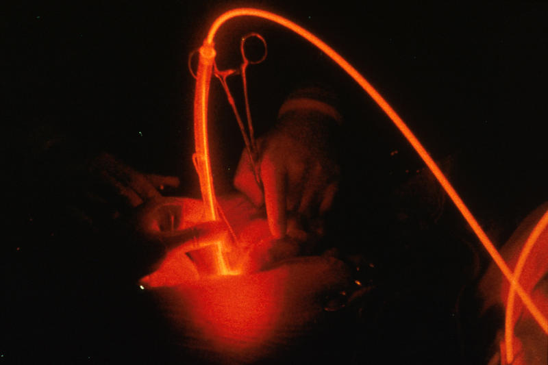
This disease can be arrested at the initial stage with the help of medication. But a person suffers a heart attack when the precautions are not taken, or when the artery is fully obstructed. That is when the surgical procedure of angioplasty is done. In angioplasty, a balloon is introduced into the artery through a guide wire and is inflated where the blockage is located. After this the stent is introduced so that it keeps the artery open.
The biodegradable stent releases a drug called everolimus which prevents irregular tissue growth.
Researches and studies that classify biodegradable as safe
Kunhiko Kosuga, who has a MD, PhD and is also the director of cardiology at Shiga Medical Center for Adults in Moriyana City, Japan, did a research on these new stents. He and his fellow researchers studied 44 men and 6 women who had undergone angioplasty and had used biodegradable stents to open up the affected arteries. They looked for various complications like clots, deaths, and other causes. The result is as follows:
◦ for the deaths associated with heart diseases, the survival rate was 98%.
◦ for death from all causes, the survival rate was 87%.
◦ there was no main cardiac problems in half the patients.
◦ Only four patients suffered heart attacks.
◦ The blood vessel involved had re-narrowed in 16% of the patients, in one year after undergoing the procedure.
◦ there were two clots that were found within the stent. One was due to the drug-infused stent close to the biodegradable one.
Countries who welcomed biodegradable stents
Nine European countries, Middle East, parts of Latin America and parts of Asia like India, Hong Kong, Philippines and Vietnam are already using these stents. In Europe, Asia-Pacific, Canada and Latin America, over 600 patients have taken part in the trial which aspires to have 1000 patients from over 100 centres present in these counties. Even Singapore has approved of these stents from 20th December, 2012.
However, doctors are still awaiting results for the long term effects on the patients
Even though the cost for manufacturing these stents is very expensive, doctors worldwide are optimistic that they will replace metallic stents eventually.
About The Author: Alia is a writer/blogger by profession. She loves writing, travelling and reading books. She contributes to Hydroxycut
Courtesy: Alia and http://www.biotechblog.org/

Life-Producing Phosphorus Carried to Earth by Meteorites


June 4, 2013 — Scientists may not know for certain whether life exists in outer space, but new research from a team of scientists led by a University of South Florida astrobiologist now shows that one key element that produced life on Earth was carried here on meteorites.


In an article published in the new edition of the Proceedings of the National Academy of Sciences, USF Assistant Professor of Geology Matthew Pasek and researchers from the University of Washington and the Edinburg Centre for Carbon Innovation, revealed new findings that explain how the reactive phosphorus that was an essential component for creating the earliest life forms came to Earth.
The scientists found that during the Hadean and Archean eons -- the first of the four principal eons of Earth's earliest history -- the heavy bombardment of meteorites provided reactive phosphorus that when released in water could be incorporated into prebiotic molecules. The scientists documented the phosphorus in early Archean limestone, showing it was abundant some 3.5 billion years ago.
The scientists concluded that the meteorites delivered phosphorus in minerals that are not seen on the surface of Earth, and these minerals corroded in water to release phosphorus in a form seen only on the early Earth.
The discovery answers one of the key questions for scientist trying to unlock the processes that gave rise to early life forms: Why don't we see new life forms today?
"Meteorite phosphorus may have been a fuel that provided the energy and phosphorus necessary for the onset of life," said Pasek, who studies the chemical composition of space and how it might have contributed to the origins of life. "If this meteoritic phosphorus is added to simple organic compounds, it can generate phosphorus biomolecules identical to those seen in life today."
Pasek said the research provides a plausible answer: The conditions under which life arose on Earth billions of years ago are no longer present today.
"The present research shows that this is indeed the case: Phosphorus chemistry on the early Earth was substantially different billions of years ago than it is today," he added.
The research team reached their conclusion after examining Earth core samples from Australia, Zimbabwe, West Virginia, Wyoming and in Avon Park, Florida.
Previous research had showed that before the emergence of modern DNA-RNA-protein life that is known today, the earliest biological forms evolved from RNA alone. What has stumped scientists, however, was understanding how those early RNA-based life forms synthesized environmental phosphorus, which in its current form is relatively insoluble and unreactive.
Meteorites would have provided reactive phosphorus in the form of the iron-nickel phosphide mineral schreibersite, which in water released soluble and reactive phosphite. Phosphite is the salt scientists believe could have been incorporated into prebiotic molecules.
Of all of the samples analyzed, only the oldest, the Coonterunah carbonate samples from the early Archean of Australia, showed the presence of phosphite, Other natural sources of phosphite include lightning strikes, geothermal fluidsand possibly microbial activity under extremely anaerobic condition, but no other terrestrial sources of phosphite have been identified and none could have produced the quantities of phosphite needed to be dissolved in early Earth oceans that gave rise to life, the researchers concluded.
The scientists said meteorite phosphite would have been abundant enough to adjust the chemistry of the oceans, with its chemical signature later becoming trapped in marine carbonate where it was preserved.
It is still possible, the researchers noted, that other natural sources of phosphite could be identified, such as in hydrothermal systems. While that might lead to reducing the total meteoric mass necessary to provide enough phosphite, the researchers said more work would need to be done to determine the exact contribution of separate sources to what they are certain was an essential ingredient to early life.

Genetic Editing Shows Promise in Duchenne Muscular Dystrophy


June 4, 2013 — Using a novel genetic 'editing' technique, Duke University biomedical engineers have been able to repair a defect responsible for one of the most common inherited disorders, Duchenne muscular dystrophy, in cell samples from Duchenne patients.


Instead of the common gene therapy approach of adding new genetic material to "override" the faulty gene, the Duke scientists have developed a way to change the existing mutated gene responsible for the disorder into a normally functioning gene. The Duke researchers believe their approach could be safer and more stable than current methods of gene therapy.
The researchers are now conducting further tests of this new approach in animal models of the disease.
Duchenne muscular dystrophy is a genetic disease affecting one in 3,600 newborn males. The genetic mutation is found on the X chromosome, of which males have only one copy. (Females, with two X chromosomes, presumably have at least one good copy of the gene.)
Patients with Duchenne muscular dystrophy cannot produce the protein known as dystrophin, which is essential in maintaining the structural integrity of muscle fibers. Over time, patients with the disorder suffer gradual muscle deterioration, which leads to paralysis and eventual death, usually by age 25.
"Conventional genetic approaches to treating the disease involve adding normal genes to compensate for the mutated genes," said Charles Gersbach, assistant professor of biomedical engineering at Duke's Pratt School of Engineering and Department of Orthopaedic Surgery and member of Duke's Institute for Genome Sciences and Policy. "However, this can cause other unforeseen problems, or the beneficial effect does not always last very long.
"Our approach actually repairs the faulty gene, which is a lot simpler," said David Ousterout, the Duke biomedical engineering graduate student in the Gersbach lab who led the work. "It finds the faulty gene, and fixes it so it can start producing a functional protein again."
The results of the Duke study were published online inMolecular Therapy, the journal of the American Society for Gene and Cell Therapy. The project was supported by the Hartwell Foundation, the March of Dimes Foundation and the National Institutes of Health.
The Duke experiments, which were carried out in cell samples from Duchenne muscular dystrophy patients, were made possible by using a new technology for building synthetic proteins known as transcription activator-like effector nucleases (TALENs), which are artificial enzymes that can be engineered to bind to and modify almost any gene sequence.
These TALENs bind to the defective gene, and can correct the mutation to create a normally functioning gene.
"There is currently no effective treatment for this disease," Gersbach said. "Patients usually are in a wheelchair by the age of ten and many die in their late teens or early twenties."
Duchenne muscular dystrophy has been extensively studied by scientists, and it is believed that more than 60 percent of patients with this type of mutation can be treated with this novel genetic approach.
"Previous studies indicate that restoring the production of dystrophin proteins will be highly functional and alleviate disease symptoms when expressed in skeletal muscle tissue," said Ousterout.
Similar approaches could be helpful in treating other genetic diseases where a few gene mutations are responsible, such as sickle cell disease, hemophilia, or other muscular dystrophies, Gersbach said.
Other members of the team were Duke's Pablo Perez-Pinera, Pratiksha Thakore, Ami Kabadi, Matthew Brown, Xiaoxia Qin, and Olivier Fedrigo. Other participants were Vincent Mouly, Universite Pierre at Marie Curie, Paris, and Jacques Tremblay, Universite Laval, Quebec.

Courtesy: http://www.sciencedaily.com/releases/2013/06/130604153946.htm